Lung Parenchymal - Nodular Lesions In The Lung Parenchyma Sesc Historial De Consultes - Lung parenchyma is the medical term used to describe the actual functioning parts of a human or animal lung.
byAdmin•
0
Lung Parenchymal - Nodular Lesions In The Lung Parenchyma Sesc Historial De Consultes - Lung parenchyma is the medical term used to describe the actual functioning parts of a human or animal lung.. A lung nodule is a term for small abnormalities seen on xray and ct scans. The most accurate way to determine if a lung disease affects this part of the lung is with a surgical biopsy. Parenchymal bands are most frequently encountered in individuals who have been exposed to. Airways, alveoli, interstitium, and vasculature. Diffuse parenchymal lung diseases are disorders that affect the interstitial of the lungthe area around the lung's air sacs.
In some cases, however, the causes remain unknown. Fibroblast activation results in the formation of fibroblastic foci at the margins of normal lung composed of dense collagen. Apical parenchymal scarring, for example, is scarring at the tip of the lung. Lung parenchyma what is lung parenchyma lung parenchyma Ct can demonstrate interstitial lung disease, emphysema associated with chronic obstructive pulmonary disease, features of left heart failure (including interstitial oedema), and changes secondary to miscellaneous conditions such as sarcoidosis.
A Gross Lesions In Lung Parenchyma Single And Coalescent Parasitic Download Scientific Diagram from www.researchgate.net Most lung nodules are benign (not cancerous). Some types of autoimmune diseases, such as rheumatoid arthritis, also can cause interstitial lung disease. In both intrinsic or extrinsic pulmonary conditions, lung volumes become reduced due to restrictions in pulmonary mechanics. Diffuse parenchymal lung diseases are disorders that affect the interstitial of the lungthe area around the lung's air sacs. Find this author on pubmed. Ct parenchymal lung changes can help to differentiate the aetiology of ph. Brody, md cincinnati children's hospital. Among the injuries considered in this article are traumatic pulmonary pseudocysts, pulmonary hematomas, major pulmonary lacerations, pulmonary contusions, and penetrating pulmonary parenchymal injuries.
Pulmonary parenchymal damage is a frequent consequence of major trauma to the chest.
Pulmonary parenchymal damage is a frequent consequence of major trauma to the chest. A lung (pulmonary) nodule is an abnormal growth that forms in a lung. Lung parenchyma is the medical term used to describe the actual functioning parts of a human or animal lung. If lung tissue is obtained, however, It includes the alveolar walls as well as the blood vessels and the bronchi. Diffuse parenchymal lung diseases are disorders that affect the interstitial of the lungthe area around the lung's air sacs. Among the injuries considered in this article are traumatic pulmonary pseudocysts, pulmonary hematomas, major pulmonary lacerations, pulmonary contusions, and penetrating pulmonary parenchymal injuries. In both intrinsic or extrinsic pulmonary conditions, lung volumes become reduced due to restrictions in pulmonary mechanics. Apical parenchymal scarring, for example, is scarring at the tip of the lung. These abnormalities may be due to a disease of the pulmonary interstitial tissue, the bronchial tree, the cardiovascular system or to abnormal alveolar filling with fluid, blood, cells or tumor, several of these etiologies possibly being concomitant. Wiki says, pulmonary fibrosis involves gradual exchange of normal lung parenchyma with fibrotic tissue. A blinded pulmonary pathologist examined the lung parenchyma focusing on four compartments: Lung parenchyma is the substance of the lung outside of the circulatory system that is involved with gas exchange and includes the pulmonary alveoli and respiratory bronchioles, though some authors include only the alveoli.
Parenchymal bands are most frequently encountered in individuals who have been exposed to. Ttp, ards, chest tube 10 yrs ago. Parenchymal scarring is scarring of the tissue in the lungs. Lung parenchyma what is lung parenchyma lung parenchyma Among the injuries considered in this article are traumatic pulmonary pseudocysts, pulmonary hematomas, major pulmonary lacerations, pulmonary contusions, and penetrating pulmonary parenchymal injuries.
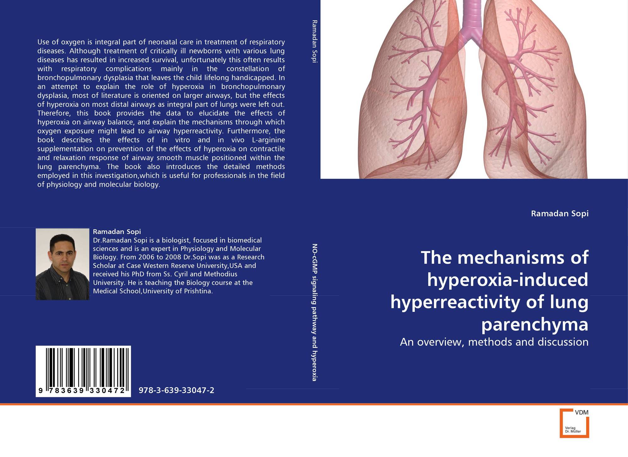
The Mechanisms Of Hyperoxia Induced Hyperreactivity Of Lung Parenchyma 978 3 639 33047 2 3639330471 9783639330472 By Ramadan Sopi from images.our-assets.com Analysis of multiple lung parenchymal abnormalities on hrct is a real diagnostic challenge. These abnormalities may be due to a disease of the pulmonary interstitial tissue, the bronchial tree, the cardiovascular system or to abnormal alveolar filling with fluid, blood, cells or tumor, several of these etiologies possibly being concomitant. Parenchymal scarring is scarring of the tissue in the lungs. Pain can arise from the parietal pleura, the major airways, the chest wall, the diaphragm, and the mediastinal structures. Parenchymal lung diseases are disorders that affect the pulmonary interstitium. Nodules may develop in one lung or both. A lung (pulmonary) nodule is an abnormal growth that forms in a lung. When active alveolitis is present, a variety of inflammatory cells (e.g., macrophages, lymphocytes, neutrophils, eosinophils, and plasma cells) infiltrate the alveolar wall.
Ct can demonstrate interstitial lung disease, emphysema associated with chronic obstructive pulmonary disease, features of left heart failure (including interstitial oedema), and changes secondary to miscellaneous conditions such as sarcoidosis.
Pain can arise from the parietal pleura, the major airways, the chest wall, the diaphragm, and the mediastinal structures. Diffuse parenchymal (interstitial) lung diseases are characterized pathologically by variable amounts of alveolitis and fibrosis. Parenchymal lung diseases are disorders that affect the pulmonary interstitium. The lung parenchyma will have a heterogeneous appearance with patchy areas of normal lung, areas of mild interstitial inflammation, fibrosis, and honeycombing. Pulmonary parenchymal damage is a frequent consequence of major trauma to the chest. The most accurate way to determine if a lung disease affects this part of the lung is with a surgical biopsy. When active alveolitis is present, a variety of inflammatory cells (e.g., macrophages, lymphocytes, neutrophils, eosinophils, and plasma cells) infiltrate the alveolar wall. The lung parenchyma and the visceral pleura are insensitive to most painful stimuli, and interference with stretch fibers tends to cause most intrapulmonary symptoms. Ct parenchymal lung changes can help to differentiate the aetiology of ph. Pulmonary edema (pe) blood transfusions. 1 dept of diseases of the thorax, gb morgagni hospital, forlì, italy. Diffuse alveolar damage (dad/ards) bleomycin, busulfan, cyclophosphamide, mitomycin, amiodarone. Review causes of opacities other than infection!
If any part of the parenchyma becomes damaged or diseased, a person's life may be at risk. Apical parenchymal scarring, for example, is scarring at the tip of the lung. In some cases, however, the causes remain unknown. Seventeen year old with fever. Lung parenchyma is the substance of the lung outside of the circulatory system that is involved with gas exchange and includes the pulmonary alveoli and respiratory bronchioles, though some authors include only the alveoli.
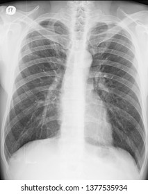
Infiltration Lung Parenchyma High Res Stock Images Shutterstock from image.shutterstock.com Once lung scarring occurs, it's generally irreversible. Pulmonary edema (pe) blood transfusions. Pulmonary parenchymal opacities other than infection alan s. A blinded pulmonary pathologist examined the lung parenchyma focusing on four compartments: Ct parenchymal lung changes can help to differentiate the aetiology of ph. Can't get over 1k on incent spirometer since. Most lung nodules are benign (not cancerous). The percentage is higher on computed tomography which can detect disease when the radiograph is normal.
Pulmonary edema (pe) blood transfusions.
Pulmonary parenchymal lymphoma generally presents as nodules, consolidative infiltrates, or peribronchovascular opacities. Once lung scarring occurs, it's generally irreversible. Most lung nodules are benign (not cancerous). The percentage is higher on computed tomography which can detect disease when the radiograph is normal. Find this author on google scholar. Can't get over 1k on incent spirometer since. The alveoli are held open by the transpulmonary pressure, or prestress, which is balanced by tissues forces and alveolar surface film forces. These abnormalities may be due to a disease of the pulmonary interstitial tissue, the bronchial tree, the cardiovascular system or to abnormal alveolar filling with fluid, blood, cells or tumor, several of these etiologies possibly being concomitant. Ct parenchymal lung changes can help to differentiate the aetiology of ph. A lung (pulmonary) nodule is an abnormal growth that forms in a lung. Review causes of opacities other than infection! Parenchymal scarring is scarring of the tissue in the lungs. Am i high risk for covid lung complications?